| CATEGORII DOCUMENTE |
| Bulgara | Ceha slovaca | Croata | Engleza | Estona | Finlandeza | Franceza |
| Germana | Italiana | Letona | Lituaniana | Maghiara | Olandeza | Poloneza |
| Sarba | Slovena | Spaniola | Suedeza | Turca | Ucraineana |
DOCUMENTE SIMILARE |
||
|
||
TERMENI importanti pentru acest document |
||
| : | ||
|
Helix aspersa Helix aspersa Helix,
the edible terrestrial snail, is often used in invertebrate zoology courses
as an example of pulmonate anatomy. Helix
pomatia is the escargot and H. aspersa is the garden snail, a
smaller species. The latter has been
introduced to the west coast of Helix is a typical, shelled, coiled, torted pulmonate snail and as such is well chosen to serve as a representative pulmonate. The anatomy of Helix and the slugs is similar except that Helix has a shell and is coiled. Because of these two differences, Helix is a more difficult dissection. The shell and coiling complicate the dissection and the body cavity is more crowded. If both are available, large slugs such as Limax, Arion, or Ariolimax are much easier to dissect. Further, slugs are easier to anesthetize in a relaxed condition. Anesthetization Helix is especially difficult to anesthetize in a relaxed and extended condition. The snail will retract into its shell when placed in almost any environment other than air or water. The older literature recommends drowning the animal by placing it in a jar of water over which there is no air space. The snail eventually succumbs in a more or less extended condition. They can also be killed and partly relaxed more quickly and humanely by placing them in 20% ethanol in water for about an hour. As is true of any snail, living or freshly killed specimens are much better than preserved. If living animals are fed a diet of dry cat food, such as Purina Main Meal, the gut will be conveniently color coded, in this case pink. External Anatomy Place a preserved or relaxed, extended, freshly killed animal in a dish of pondwater and examine its exterior. Shell Helix is coiled and torted. Its shell is thin, fragile, fat, and globular. It is brown with darker brown spiral stripes and is covered by a thin proteinaceous periostracum (Fig 1). Figure 1. The shell of the garden snail, Helix aspersa, with the animal fully retracted. gastrop75L.gif
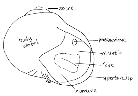
The shell, like that of any coiled gastropod, is a hollow cone spiraled around a central axis, the columella. The columella cannot be seen until you break the shell. Each complete turn of the cone is a whorl. Almost all of the shell of Helix is in the last whorl, which is known as the body whorl. Most of the body of the snail resides in the body whorl. There are a few much smaller whorls above the body whorl and these together comprise the spire. The spire of Helix is small and sits atop the body whorl. The body whorl opens to the exterior vial a large opening, the aperture, from which the animal extends its head and foot. There is no operculum, the absence of which is characteristic of pulmonates. The edge of the aperture is the aperture lip. Examine the visible parts of the soft anatomy of your specimen. The body consists of a head, foot, and visceral mass. The head and foot can be extended from the aperture and should be visible now. The visceral mass always remains in the shell and cannot be extended. Remove the animal from its shell by breaking bits of the shell away with a strong pair of forceps or a C-clamp. Begin at the aperture and work your way up the body whorl. Be careful you do not damage and of the soft tissues. The animal is attached to the shell at only one point by the columellar muscle. This strong white muscle has its origin on the columella well inside the aperture on the right side of the animal. It inserts on the posterior foot. Take this opportunity to find the columella, which is the central axis of the shell. Orientation Clean away the pieces of the shell and immerse the snail in water again. Find some familiar landmarks such as the head, foot, and visceral mass and orient yourself. The animal looks quite different without its shell and you may be temporarily disoriented. Spend a little time finding the major axes. Orientation of coiled gastropods is a little difficult due to their asymmetry. The animal is elongated along two axes, the anteroposterior and the dorsal ventral. The head and foot lie on the anteroposterior axes and are elongated along it. This axis is the axis of symmetry of the head and foot, which are bilaterally symmetrical. Neither head nor foot is affected by coiling or torsion. The visceral mass, on the other hand, sits on the dorsal surface of the foot and is elongated along the dorsoventral axis. It coils up into the spiral shell and is asymmetrical. Find anterior, posterior, dorsal, ventral, right, and left. Do this using the head and foot while ignoring the visceral mass. Remember that the head is anterior, the opposite end of the foot is posterior. The sole of the foot is ventral and the visceral mass is dorsal. Head The head is at the anterior end of the animal and is well developed (Fig 2). It bears the mouth at its anterior end. The mouth is flanked by large fleshy lips. The head bears two pairs of retractile sensory tentacles at its anterior dorsal corners. The more dorsal and posterior cephalic tentacles, are the larger and each bears an eye at its tip. The much smaller, more ventral and anterior oral tentacles have no eyes. The common gonopore of the hermaphroditic reproductive system is located on the right side of the head. It is slightly posterior to the right cephalic tentacle. The foot is a long, wide, muscular organ with a smooth flat sole. The sole fits against the substratum and creeps along it. A mucus-secreting pedal gland opens between the head and the foot and secretes lubricating mucus onto the substratum in the path of the foot. Place the snail in the pan with the sole of the foot against the wax, as if it were crawling, and anchor the foot to the wax with two # 1 insect pins. Find the visceral mass if you have not already done so. It is the large, coiled mass of tissue sitting on the dorsal surface of the foot, posteriad the head. It was entirely hidden by the shell but is now visible. It is everything other than the head and foot. Mantle and Mantle Cavity The mantle is the body wall of the anterior dorsal surface of the visceral mass. It is immediately posteriad the head. The mantle secretes the shell. The mantle is folded to form a deep recess, the mantle cavity, posterior to the head on the dorsal side of the animal. The mantle cavity of pulmonates is a large air space enclosed by the mantle. The edge anterior of the mantle of Helix forms a conspicuous collar, or skirt, which you see as a high fleshy ridge encircling the dorsal and lateral body between the head and visceral mass (Fig 2). In life the skirt fits around the lip of the aperture of the shell and seals it. Figure 2. Helix aspersa viewed from the right side. The shell has been removed. gastrop76L.gif
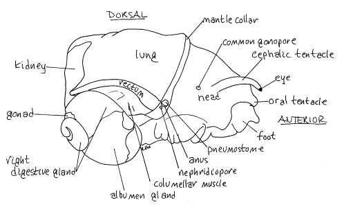
In most molluscs the gills are located in the mantle cavity but pulmonates have no gills. Instead the interior of the mantle cavity is vascularized to function as a lung for respiration in air. In the prosobranch gastropods there would be a large anterior opening into the mantle cavity between the mantle skirt and the body but in pulmonates this opening is reduced to a pneumostome on the right side on or just below the edge of the collar (Fig 2). Although the opening to the mantle cavity is reduced, the cavity itself is very large and occupies most of the region at the base of the visceral mass. Its dorsal wall, which is the mantle, is very thin and translucent (in life). The large anus lies beside the pneumostome on its posterio-ventral border. Urinary wastes exit via a small nephridiopore in the ventral right corner of the pneumostome. Many large organs are visible on the surface of the visceral mass of living specimens. They are not apparent in preserved animals. If you have a living or fresh animal, you should find them now as they make useful landmarks. Through the thin roof of the mantle cavity you can see the large, white, triangular kidney at the posterior end of the mantle cavity. The translucent, white heart lies on the left side of the kidney. The rectum, whose color reflects the diet, lies along the far right border of the mantle cavity. The white columellar muscle lies to the right of the rectum. The base of the body whorl is occupied by the large, brown left digestive gland. A segment of the intestine can be seen imbedded in it. As you follow the coil of the visceral mass, the white albumen gland follows the left digestive gland. The remainder of the coil is occupied by the right digestive gland and the pale cream colored hermaphroditic gonad, the ovotestis. Use fine scissors to open the mantle cavity by making a longitudinal dorsal incision from the pneumostome to the posterior end of the cavity along the right side of the mantle. The cut will pass between the right side of the large bulging kidney and the tubular rectum. Do not cut either of these organs. The incision will be a long one as the mantle cavity is very deep. Make another cut transversely from the pneumostome to the left side of the mantle along the posterior edge of the collar. Turn and cut posteriorly along the left side of the mantle to the posterior end of the mantle cavity. This cut will pass to the left of the heart and kidney. Do not cut anything other than the thin, translucent mantle. Avoid the organs in it and those below the cavity. Deflect the mantle, which is still attached to the body along its posterior edge. The mantle cavity is now open and accessible. Figure 3. Dorsal view of Helix aspersa with the mantle cut and deflected posteriorly. gastrop77L.gif
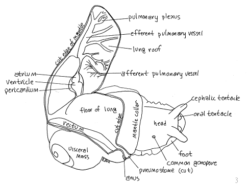
The space revealed by deflecting the mantle is the mantle cavity, or lung. Its roof is, of course, the mantle and its floor is the dorsal body wall, which is thin and transparent in this region (Fig 3). Some of the internal organs can be seen through the floor of the mantle cavity. You may study them later. Note that there is no gill in the mantle cavity as there would be if this were a prosobranch. Look at the inside surface of the mantle. The large creamy white kidney bulges from the mantle into the mantle cavity and you can now get a good look at it (Fig 3). The transparent pericardium lies against the left side of the kidney, on the mantle (Fig 3). In living or freshly killed specimens the heart is clearly visible within it but in preserved material the heart will not be visible until the pericardium is opened later in the exercise. The heart consists of an anterior atrium and a slightly larger, opaque, posterior ventricle. A conspicuous complicated pulmonary plexus of blood vessels on the mantle drains into the anterior end of the atrium by a large efferent pulmonary vessel (Fig 3). A large aorta exits the posterior end of the ventricle. It immediately penetrates the dorsal body wall and you will not see it now. The tubular rectum runs along the right edge of the mantle cavity at the line of junction between the roof and floor of the mantle cavity (Fig 3). It begins at the posterior end of the cavity, where it emerges from the visceral mass, and runs anteriorly to end at the anus lying on the lower right edge of the pneumostome. The ureter leaves the kidney and runs along the outside (right) edge of the rectum. It empties at the pneumostome via the nephridiopore. Internal Anatomy Now open the body cavity (which is a hemocoel) with a longitudinal, middorsal incision, through the body wall. Make this cut with fine scissors, from the mouth posteriorly, through the collar, and along the floor of the mantle cavity. Be careful that you cut no deeper than the body wall. The body wall of the head is thick and tough but that of the floor of the mantle cavity is very thin. The collar is also thick. Do not damage the internal organs, most of which belong to the digestive and reproductive systems. Pin the body wall aside. The cavity you have revealed is the hemocoel and it is tightly packed with organs. In life it would be filled with blood (hemolymph). Find the following structures to use as landmarks. In the head find the buccal mass. This large, ovoid mass of muscle contains the pharynx and radula. You will open it later. The large, dark (its color depends on that of the last meal), tubular crop lies on the left and extends the length of the hemocoel. The complex, mostly white reproductive system occupies all the space on the right. Blood Vascular System Relocate the pericardium in the roof of the mantle and use fine scissors to open it to reveal the atrium and ventricle more clearly. The atrium is anterior and the ventricle is posterior. The efferent pulmonary vessel drains the pulmonary plexus into the anterior end of the atrium. Find the aorta exiting the ventricle and trace it through the body wall into the hemocoel. It leads to an extensive system of vessels that extend throughout the hemocoel to supply its organs. Trace some of the vessels to their targets if you wish. We will not make a detailed study of the blood vascular system in this exercise. Any study of this system should be done now, before the vessels are destroyed. Free the reproductive system from its membranous connections with the dorsal body wall and crop and move it to the right, out of the way. Nervous System The narrow, tubular esophagus exits the buccal mass and turns to the left to connect with the much wider crop. The brain is a large circumenteric nerve ring around the esophagus just posterior to the posterior buccal mass. The nerve ring is enclosed in a sheath of connective tissue. The sheath is a very large flat band that is easily found. The ganglia and connectives of the nerve ring are inside the sheath and, in preserved specimens, cannot be seen without further dissection. The nerve ring includes a pair of large dorsal cerebral ganglia which are connected to each other by a short cerebral commissure passing across the midline. A large connective exits each cerebral ganglion and passes ventrally and posteriorly to a cluster of ganglia ventral to the esophagus. Included in this cluster are the pleural, pedal, parietal, and visceral ganglia. Many nerves exit the ring. The large cephalic aorta can be seen penetrating the ventral cluster of ganglia on its way to the head. The optic nerve from the cephalic tentacles can be seen entering the ring. Figure 4. Dorsal dissection of Helix aspera with the mantle cavity and hemocoel open. gastrop78L.gif
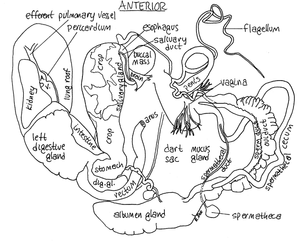
Musculature The columellar muscle originates on the columella of the shell and extends from there into the body of the snail where it divides into several long muscles which run along the floor of the hemocoel. Most of these are long, white, flat, and straplike and lie under the reproductive organs and crop. Find the buccal retractor muscles. These two branches of the columellar muscle pass from the origin at the columella through the nerve ring to insert on the buccal mass. Their action is to withdraw the buccal mass and head into the shell and body. Two other slips of the columellar muscle, the tentaclular muscles, run to the two cephalic tentacles. Their action is to withdraw the tentacles. Near the tentacles they are dark grey or black. They do not pass through the nerve ring. Yet another pair of muscles runs to the oral tentacles and there is also a penis retractor muscle. The remainder of the columellar muscle fans out into numerous paired slips to all regions of the foot. These are the pedal retractor muscles and their action is to withdraw the foot into the shell. Digestive System Relocate the buccal mass in the center of the head. Use fine scissors to open it with a longitudinal, middorsal incision beginning at the mouth. Be careful that you do not cut the nerve ring which encircles its posterior end. This incision will reveal the lumen of the anterior gut. The mouth opens into a small anterior buccal cavity equipped with a single, dark, heavy, toothed jaw . The jaw is located dorsally over the opening of the mouth into the buccal cavity. You cut through it when you opened the buccal mass. Most of the interior of the buccal cavity is the pharynx. The radula lies on the floor of the pharynx. The radula is a wide, golden brown, thin, transparent sheet of tough tissue which bears numerous tiny teeth. The radula is situated on the dorsal surface of the muscular odontophore, which contributes to the floor of the pharynx. Remove the radula and make a wetmount of it for study with the compound microscope. A short, narrow esophagus exits the posterior end of the pharynx, passes through the nerve ring, turns to the left and widens to become the anterior end of the crop (Fig 4). The crop is a large, thin-walled storage organ extending the entire length of the hemocoel. The snail's last meal may be visible inside it. A pair of diffuse, white (in life) salivary glands partially cover the walls of the anterior end of the crop (Fig 4). In preserved specimens the salivary glands are about the same beige color as the crop. Each gland drains to the pharynx via a salivary duct. The ducts pass through the nerve ring. Follow the crop posteriorly, past the origin of the columellar muscle. In this region it is obscured by the large, white albumen gland and the two dark brown digestive glands. The right digestive gland is in the upper, small whorls of the visceral mass and, in fact, accounts for most of the volume of the upper whorls. The pale cream ovotestis is also in the upper whorls located along the inside curve of the spiral. The large white albumen gland of the reproductive system is also present in the spiral of the visceral mass but is lower down, near the base. It is in the middle region of the spiraled visceral mass. The left digestive gland lies at the base of the visceral mass and extends into the mantle roof to lie against the kidney. Use fine scissors to cut the connective tissue necessary to separate the albumen gland from the left digestive gland. Cut posteriorly along the left side of the rectum, following the rectum until it disappears from view into the visceral mass. This will expose most of the albumen gland. Be sure you do not cut into the gland. Cut the membranous connective tissue holding the albumen gland to the crop but do not cut any organs. When all these connections are severed, grasp the albumen gland gently with forceps and pull it out of the pocket it fills in the middle whorls of the visceral mass. Do not break or damage the convoluted, white hermaphroditis duct which runs from the ovotestis to the albumen gland. You cannot see all of the ovotestis yet. The hermaphroditic duct is lost from view under some of the branches of the columellar muscle. Between the two digestive glands the crop expands to become the stomach, into which the digestive glands open. The stomach narrows to become the intestine which is also embedded in the left digestive gland. It makes a large loop as it passes through the gland. The intestine emerges from the digestive gland as the rectum, which you have already seen. The rectum runs anteriorly along the right side of the mantle cavity to open at the anus. Reproductive System Pulmonates are hermaphrodites with internal fertilization. Both conditions require modifications of the reproductive system and that of Helix is one of the most complicated you will encounter. Fortunately almost all parts of it lie free in the hemocoel and are relatively easily found and identified. The protandric (male first then female) ovotestis is located at the top of the visceral mass high in the spire (Fig 2). It is surrounded by the digestive gland and is pale creamy-white (in life) or dark brown (preserved). In preserved specimens the ovotestis is darker than the digestive gland whereas the opposite is true of fresh material. The degree of pigmentation of pulmonate gonads depends on the reproductive condition of the individual. A small portion of the gonad can be seen (barely) without further dissection on the surface of the visceral mass in the groove between the smallest whorls. It is almost completely surrounded by the right digestive gland. You will have to dig into the right digestive gland to see more of the ovotestis but you should not do so at this time. Relocate the albumen gland and separate it from its surrounding tissues if you have not already done so. The albumen gland secretes a nutritive material around the fertilized egg. Its size varies with changing reproductive condition but it can be quite large. A small, but conspicuous (in life), white, convoluted hermaphroditic duct extends from the ovotestis to the albumen gland. Autochthonous spermatozoa are stored in the hermaphroditic duct. Fertilization occurs in a fertilization chamber embedded in the albumen gland (at the point where the hermaphroditic duct, albumen gland duct and spermoviduct join.) Anteriorly the albumen gland is continuous with a very thick and conspicuous spermoviduct or common gonoduct (Fig 4). This is a combined sperm duct and oviduct running side by side. The large sacculated side of the duct is the oviduct whereas the smaller bright white inner margin is the sperm duct. The lumina of the two ducts are not completely separated from each other. The oviduct secretes a calcareous shell around the fertilized egg as it passes downstream. The reproductive plumbing is complicated by the presence of two other ducts partially associated with the exterior of the spermoviduct. These are both associated with the spermatheca, a small spherical sac attached to the wall of the upper end of the spermoviduct. The spermatheca stores allochthonous sperm until needed for fertilization. The spermathecal duct is much easier to find than the spermatheca itself (Fig 4). It exits the spermatheca and runs anteriorly along the wall of the spermoviduct but is not functionally associated with it. Similarly, a blind tube, the spermathecal caecum, arises on the other side of the spermoviduct and follows a twisting course anteriorly eventually to join the spermathecal duct. The sperm duct is less obvious than either of these and appears as a white band in the wall of the spermoviduct, not as an independent duct. As the spermoviduct nears the head it divides into a separate oviduct and sperm duct. The oviduct joins the distal end of the spermathecal duct which then joins the large, pyriform, muscular dart sac to form the short vagina (Fig 4). The dart sac secretes and stores a calcareous dart which is stabbed into the snail's partner, presumably to stimulate it to copulate. A pair of large, much branched mucus glands arise from the dart sac. The mucus glands secrete mucus for lubrication. The sperm duct exits the spermoviduct as a discrete independent duct and turns medially, passes over the base of the dart sac and vagina and under the right cephalic tentacle retractor muscle, then expands in diameter to become the penis (Fig 4). During copulation, the penis is inserted into the gonopore and vagina and delivers sperm to the partner. At the point where the penis begins to expand it is joined by a long, slender, blind diverticulum, the flagellum. Spermatozoa from the sperm duct are packaged into spermatophores by the flagellum. The penis joins the vagina to form a short common chamber which opens to the exterior through the common gonopore. You saw the gonopore earlier when you studied the right side of the head. Behavior Observe the behavior of a living animal in a dish or terrarium. Note how the animal crawls on the sole of its foot. Waves of muscular contraction move from posterior to anterior along the foot to move the animal. Such waves are said to be prograde since they move in the same direction as the animal. Retrograde waves move in the opposite direction. Watch the sole of the foot of a snail through the glass as the snail crawls across the walls of a terrarium or glass dish. Are the waves retrograde or prograde? References Bullough. W. S. 1958. Practical
Invertebrate Anatomy (2nd ed). MacMillan, Hyman,
L. H. 1967. The Invertebrates, Mollusca,
vol. VI. Fretter, V. & J. Peake. 1975. Pulmonates
1. Functional Anatomy and Physiology. Academic Press, Rowett, H.G.Q. 1957. Dissection Guides V. Invertebrates. | |||
|
Politica de confidentialitate | Termeni si conditii de utilizare |

Vizualizari: 4509
Importanta: ![]()
Termeni si conditii de utilizare | Contact
© SCRIGROUP 2026 . All rights reserved