| CATEGORII DOCUMENTE |
| Bulgara | Ceha slovaca | Croata | Engleza | Estona | Finlandeza | Franceza |
| Germana | Italiana | Letona | Lituaniana | Maghiara | Olandeza | Poloneza |
| Sarba | Slovena | Spaniola | Suedeza | Turca | Ucraineana |
ENCHONDROMATOSIS-OLLIER
DISEASE-CASE REPORT
O Adam1,ES Boia2, Ramona Mandrusca3
Childrens
Hospital Louis Turcanu-Department of Pediatric Surgery,
2University of Medicine and Pharmacy Victor
Babes
ABSTRACT
Enchondromas are common intraosseous, usually benign cartilaginous tumors, that develop in close proximity to growth plate cartilage. When multiple enchondromas are present, the condition is called enchondromatosis also known as Ollier disease. Clinical manifestation often appear in the first decade of life. Ollier disease is characterized by an asymmetric distribution of cartilage lesions and these can be extremely variable.Clinical problems caused by enchondromas include skeletal deformities, limb-length discrepancy and the potential risk for malignant change to chondrosarcoma. The condition in which multiple enchondromatosis is associated with soft tissue hemangiomas is known as Maffucci syndrome.The diagnosis is based on clinical and conventional radiological evaluations.Histological analysis has a limited role and is mainly used if malignancy is suspected.
Key words:Ollier disease, enchondromatosis, multiple enchondromatosis, dyscondroplasia
Definition
Enchondromas are common benign, usually asymptomatic cartilage tomors, which develop in the metaphyses and may become incorporated into the diaphyses of long tubular bones, in close proximity to growth plate cartilage.
Enchondromatosis or Ollier disease is defined by the presence of multiple enchondromas and characterized by an asymmetric distribution of cartilage lesions that can be extremely variable in terms of size, number, location, evolution of enchondromas, age of onset and of diagnosis, requirment for surgery.
The condition in which multiple enchondromatosis is associated with soft tissue hemangiomas
known as Maffucci syndrome.
Epidemiology
The estimated prevalence of Ollier disease is 1/100.000.Maffucci disease is even rare.
Solitary endochondromas are most commonly
discovered between 20 and 40 years of age but
Ollier disease tends to present before 10 years old.
Boys are affected twice as often as girls.
Etiology and pathogenesis
Endochondral bone ossification is a highly regulated process, which requires the progression of
Undifferentiated mesenchymal cells into hypertophic chondrocytes and the subsequent replacement of a cartilaginous matrix by mineralized bone.Enchondromas develop n the methaphysis of long tubular bones in close proximity to the growth plate.Consequently, it was
proposed that they result from abnormalities in signaling pathways controlling the proliferation and
differentiation of chondrocytes, leading to the development of intraosseous cartilaginous foci.
Genetics
Ollier disease and Maffucci syndrome are usually non-familial disorders and both disorders thus appear to occur spontaneously and are not inherited.The irregular distribution of the lesions
in Ollier disease strongly suggest that it is a disor-
der of endochondral bone formation that occurs due
to a post-zygotic somatic mutation that result in mo
saism
Although an identical heterozygous mutation in the PTHR1 gene has been identified other mutations in this gene were identified in additional patients with this disorder.These studies suggest that the cause of
Ollier disease is heterogenous and raise the possibi-
lity that two or more genetic mutations are required to develop the disease.
Additional mutational events may underly progression from enchondromas to tumors.
Histopathology
Macroscopic examination of enchondromas usually
shows multiple oval-shaped or round cartilaginous nodules in osseous portions of bone.The individual
nodules are limited at their periphery by woven or
lamellar bone and are separated from each other by
intertrabecular marrow spaces.
The cartilaginous tumor matrix is usually solid,with
myxoid changes, which manifest as frayings of the matrix.Enchondromas are characterized by the pre-
sence of a striking heterogeneity and diversity in the degree of cellularity and chondrocyte phenoty-
pe.This heterogeneity depends to some extent on factors such as localization and the patients age.
In part, due to this important cellular heterogeneity
the distinction between benign enchondromas and
malignant chondrosarcomas by histochemical cri-
teria is difficult.The histological criteria for malignancy that are used for conventional chondro-
sarcoma can not be used in Ollier disease because of the increased cellularity and therefore the distinction between enchondroma and grade I
chondrosarcoma in the context of enchondroma-
tosis is extremely difficult or even impossible.
Classification
There are six types of enchondromatosis but three are more common:
-In Ollier disease there are multiple enchondromas that are mostly unilateral or une-
venly distributed throughout the metaphases of the long bones , sparing the cranium and spine.
-In Maffucci syndrome the enchondromas occur with multiple cutaneous hemangiomas that
appear as soft tissue calcifications or phleboliths on
x-ray.
-If there is symmetrical involvement through-
out the body including the cranium, hands and feet
it is known as generalised enchondromatosis.
Clinical description
Clinical manifestations in Ollier disease often appear in the first decade of life and usually start
with the appearance of palpable bony masses on a finger or a toe, an asymetric shortening of an extremity with limping,osseous deformities asso-
ciated or not with pathologic fractures.Upon physical examination, enchondromas present
on the extremities are usually visible as masses
embedded within phalanges, metacarpal and me-
tatarsal bones.The masses increase in size as the
child grows along with asymmetrical shortening of a limb and either genu varus or genu valgus deformities. Varus deformity is very common.
Enchondromas frequently affect the long tubular
bones, particularly the tibia, the femur and/or the
fibula;flat bones, especially the pelvis, can also be
affected.The lesions are usually asymetrically dis-
tributed exclusively or predominantly affecting one
side of the body.
Affected bones are often shortened and deformed.
Bone shortening may be the only clinical sign of the disease and these bone shortenings are often associated with bone bending and curving and may lead to limitation in articular movement.
Forearm deformities are frequently encountered and
these are similar to those observed in hereditary multiple exostosis(HME).Ulnar shortening is usually more relevant than shortening of the radius.
The trunc is usually not affected except for rib en-
chondromas and scoliosis resulting from pelvis imbalance.
In childhood, the lesions are subjected to pathologic fractures.
Diagnostic methods
The diagnosis of Ollier disease is based on clinical and conventional radiological evaluations.
Histological analysis has a limited role and is mainly used if malignancy is supected.
Additional investigations such as scintigraphy,
Ultrasound, magnetic resonance imaging(MRI)
are not useful for establishing the diagnosis and they are indicated for the evaluation and survei-
llance of lesions that become symptomatic(pain,
increase in size).
Biopsy of suspicious lesions may be required.
Radiography
Enchondromas are rarely observed at birh,although
The lesions are most likely already present.X-ray
Show multiple, radiolucent, homogenous lesions who run parallel with long bone axis. The lesions usually calcify with time and become diffusely punctated or stippled, a light trabeculation may be
visible.Enchondromas are frequently assembled as
clusters, thus resulting in the metaphyseal wide-
ning.When localized at the bone border, the enchondromas produce a typical notch-like image.
The lesions are almost exclusively localized in the metaphysis of long bones and in the small bones of
the hands and feet. They are initially localized close to growth plate cartilage and then migrate progressively towards the diaphysis.The epiphyseal region next to an affected metaphysis may show irregularities.In the hands, the lesions almost never affect all metacarpal bones and phalanges.
Signs of malignant transformation should be looked for, as it is a major complication of enchondromato-
sis.These signs includes cortical erosion, extension of the tumor into soft tissues and irregularity or indistinctness of the surface of the tumor.
Enchondromas tend to be well circumscribed and to show a uniform pattern of mineralization, whereas chondrosarcomas show poor demarcation and the presence of unmineralized parts.
Differential diagnosis
The differential diagnosis may include:
-Hereditary multiple endochondromatosis-HME
Is an autosomal dominant disorder characterized by multiple bone tumors capped by cartilage, that occur mostly in the metaphyses of long bones.
-Other rare forms of chondromatosis which
include metachondromatosis,spondyloenchondro-
sia and genochondromatosis type I and II
-Polyostotic fibrous dysplasia
-Diaphyseal aclasis
-Kaposi sarcoma
-Klippel-Trenaunay-Weber syndrome
Treatment
There is no medical treatment for Ollier disease.
Surgery is indicated in case of complications-patho-
logical fractures, growth defect, malignant transfor-
mation.
Complications
As well as the problem of asymmetrical growth there can be pathological fracture and malignant change as chondrosarcoma and osteosarcoma.
In Ollier disease about 25% of patients will have had malignant change by the age of 40.
Prognosis
The prognosis for Ollier disease is difficult to assess.As in generally the case, forms with an early
onset appear more severe.Research has shown that patient with numerous lesions may have a better
prognosis than patients with localized cartilaginous changes, which may induce major shortening of a
lower extremity and thus limb asymmetry.
After puberty, the enchondromas typically stabilize as cartilage is replaced by bone.
The reported incidence of malignant trandformation is variable and estimated to occur in 5-50%.
Case report
We present a case of a six year old boy that was admitted in our department four years ago for pain and limping in the right lower extremity.
After an x-ray examination, the diagnosis of bone cyst of the proximal right femur was established.
A biopsy of suspicious lesions was performed and
the histopathological diagnosis was mistakenly established as aneurysmal bone cyst.
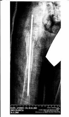
As intraoperatory event we report a pathological
fracture of the right femur occured at the lesions level and a intramedually rod was inserted followed
by casting.Postoperative period was uneventful.The images are shown bellow.
The patient was followed up clinicaly and radiolo- gicaly at two months in the next six months and afterwards every one year.The rod was removed
after six months without visible shortening of the right lower limb, but with an persistent mild limping.In the following period, the patient accused minor pain at the lower limb level and also right upper limb and foot pain were associated.
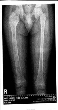
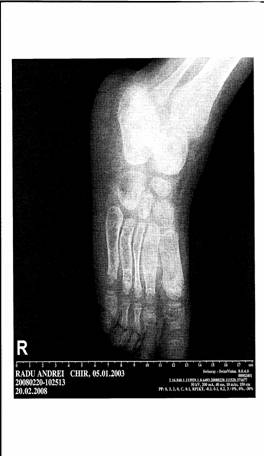
At the age of four, an x-ray of the lower and upper limb and cranium were performed and revealed multiple radiolucent, homogenous lesions with an oval shape and well defined slightly thickened bony margin-enchondromas like-localized at the superior metaphyseal and diaphyseal regions of the right femur , right tibia and at the II, III,IV, V metatarsals regions(see the images bellow).No pathologicals images at the upper limb and at the cranium level were found.
A biopsy of suspicious lesions from the right tibia was performed and the diagnosis was confirmed by the histopathological exams both in our laboratory and in Centre de Pathologie from Montpellier in France.Postoperative period was uneventful.
The follow up was continued .Two years later,in 24.04.2009, the patient was admitted in our department with a pathological fracture of the right femur following a minor injury caused by a fall from the same level.An open reduction and nailing were performed and a new biopsy was prelevated, the Ollier disease being confirmed again.
At the present time, the patient is imobilized and monitorized every two months.
Prevention
Despite the universal acceptance that Ollier disease carries a high risk of malignant change the litera-
ture data about systematic screening for early diagnosis are very poor. One paper statedWe concluded that periodic surveillance of the brain and abdomen for occult malignant lesions is indicated in patients who have enchondromatosis(12)-but failed to be more specific about the recommended frequency of screening.Another statedIt is associated with an increased risk of malignancy including intracranial chondrosarcomas.Early diagnosis is crucial;screening patients with Ollier disease is recommended.(13)Again there is no guidance as to frequency or technique.It did state that the treatment of choice for intracranial cartilaginous tumors is complete surgical excision, but this is often dificult to achieve.An alternative treatment is proton-beam therapy.
Conclusions
Ollier disease is a extremely rare, non-hereditary skeletal condition.There is no medical treatment for this disease, the only treatment is surgery in case of complications. Most of the enchondromas stabilize after puberty as cartilage is replaced by bone,but around 25% of the cases will suffer malignant transformation by the age of 40.
We recommend clinicaly follow up every year until after puberty, when ossification is completed
and long term follow up until the age of 40, every one or two years for early diagnosis of malignant change.
References
1.Maroteaux P,Le Merrer M:Les maladies osseuses de lenfant.Paris:Medecine-Sciences,Flammarion;2002
2.Unni KK:Cartilaginous lesions of bone.J Orthop Sci2001,6:457-472
3.Whyte M:Acquired Disorders of Cartilage and Bone.Washington DC; American Society for Bone and Mineral Research; 2003
4.Gabos PG,Bowen JR:Epiphyseal-metaphyseal enchondromatosis. A new clinical entity.
J Bone Joint Surg Am 1998,80:782-792
5.Loder RT, Sundberg S,Gabriel K,Mehbod A,Meyer C:Determination of bone age in children with cartilaginous dysplasia. J Pediatr Orthop 2004
6.Kronenberg HM:Developmental regulation of the growth plate. Nature 2003,423:332-336
7.Bovee JV,van Roggen JF,Cleton-Jansen AM,Taminiau AH,van der Woude HJ,Hogendoom PC:Malignant progression in multiple enchondromatosis:an autopsy-based molecular ge-netic study. Hum Patho2000, 31:1299-1303
8.Sandberg AA:Genetics of chondrosarcoma and related tumors. Curr Opin Oncol 2004,16:342-354
9.Chew DK,Menelaus MB, Richardson MD;Olliersdisease:varus angulation at the lower femur and its management.J Pediatr Orthop. 1998 Mar-Apr;18(2):202-8
10.Silve C, Juppner H;Ollier disease. Orphanet J Rare Dis.2006Sep22;1:37
11.Watanabe K,Tsuchiya H, Sakurakichi K et al;Treatment of lower limb deformities and limb-length discrepancies with the external fixator in Ollier disease .J Orthop Sci 2007 Sep;12(5):471-5.Epub 2007 Sep 28
12.Schwartz HS,Zimmerman NB, Simon MA,et al; The malignant potential of enchondromatosis. J Bo-ne Joint Surg Am 1987 Feb;69(2):269-74
13.Liu J,Hudkins PG,Swee RG,et al;Bone sarcomas associated with Ollier disease .Cancer.1987 Apr 1;59(7):1376-85
![]()
|
Politica de confidentialitate | Termeni si conditii de utilizare |

Vizualizari: 1633
Importanta: ![]()
Termeni si conditii de utilizare | Contact
© SCRIGROUP 2025 . All rights reserved